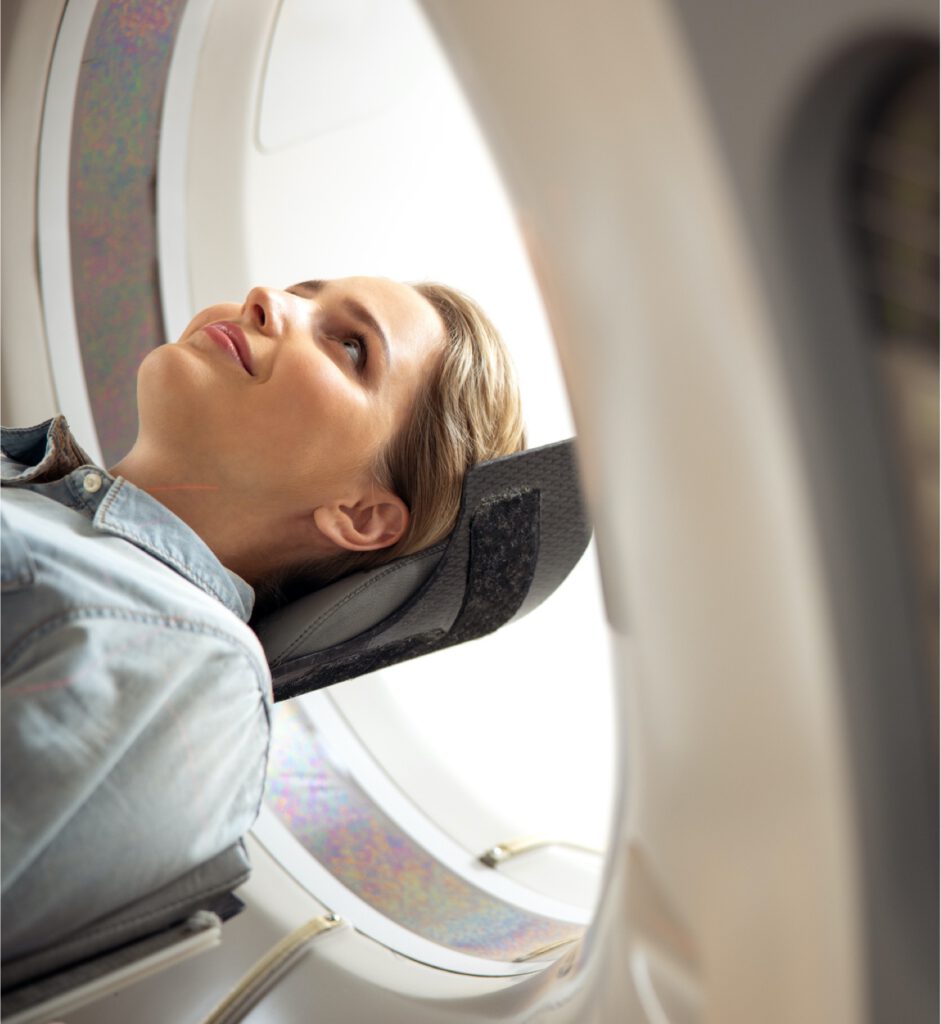
Neuroimaging

Neuroimaging experiments (fMRI, structural MRI)
Neuroimaging experiments (fMRI, structural MRI) are conducted using MRI-scanners near the clinical study site. Scanner time is rented by PI PharmaImage on-site (in the proximity of the CRO or hospital where the clinical study is conducted), PI PharmaImage provides MR-sequences and staff (technical assistants/physicians) are trained on-site if required. Imaging data analyses (e.g. event-related/task-related, resting state fMRI, volumes of hippocampus or cholinergic nuclei) are conducted remotely in Berlin. Drug challenge imaging paradigms (ketamine/nicotine) are also established. These paradigms allow testing of novel compounds whether they counteract or mimick the brain function resulting from the challenge drug. Simultaneous fMRI/EEG experiments combining the sensitivity of EEG with spatial accuracy of MRI are possible, too.
Additional physiological measurements (ECG, skin conductance), tailored stimulation paradigms (e.g. cognitive, photic stimulation) can also be provided.
All electrophysiological or neuroimaging data are transferred securely from the study site to Pharmaimage in Berlin where data analyses are conducted.
For more information see: Imaging & Electrophysiology Biomarkers
Hardware
The Pharmaimage hardware below can be delivered to the clinical study site (e.g. CRO):
Simultaneous fMRI/EEG investigations:
- 1 complete MR-compatible EEG-recording BrainAmp MR Plus system (32 channels) with 2 recording dongles, 2 laptops, 4 BrainCaps MR, 2 syncboxes
Endpoints/software standard packages:
Imaging endpoints (fMRI):
- Resting state, block design, event-related
- ROI, whole-brain voxelwise
- Functional connectivity analyses (seed-based, independent component analysis incl. default mode network analyses)
- Additionally requested parameters or MR sequences (e.g. perfusion) are possible
Software: Statistical Parametric Mapping (SPM12)
Imaging endpoints (structural MRI):
- Sequences: MPRAGE, T2-SPACE, T2 (High-Res)
- Whole-brain voxel-based morphometry, volume-based incl. subfield analyses (e.g. hippocampus, Ncl. Basalis Meynert), surface based analyses
Software: Freesurfer, DARTEL Toolbox
Functional Magnetic Resonance Imaging
Functional magnetic resonance imaging (fMRI) is increasingly used in CNS drug research. As a functional biomarker, fMRI is frequently applied when it is the aim to detect local drug effects, particularly in subcortical circuits of the brain.
Perfusion imaging (Arterial Spin Labeling) also is of tremendous value, in particular to assess brain perfusion in elderly patients with (early) vascular dementia and to evaluated whether and how a particular drug affects brain perfusion. We offer:
- Study set-up and fMRI data acquisition incl. QC/monitoring (and SOP for multicenter data acquisition).
- fMRI task conditions: resting (default mode network), N-back, verbal memory, oddball, reward etc.
- Data analysis: whole brain voxelwise, region-of-interest (ROI), seed-based functional connectivity and independent component analyses using the FSL/SPM8 software packages
- Arterial Spin labeling (ASL): Pulsed ASL sequence with echoplanar readout and online calculation of CBF (cerebral blood flow) maps.
- Pharmaco-fMRI: established subanesthetic ketamine challenge, nicotine challenge
- Reference data from N = 100 clinically stable schizophrenia patients and > N = 100 subjects with MCI (Mild Cognitive Impairment) are available (in addition to N = 1000 healthy control subjects).
- Special offer: Simultaneous fMRI/EEG. e.g. for EEG-informed functional connectivity analyses without the need to define a priori an anatomical seed. For instance, EEG information (e.g. vigilance staging during the imaging session can be used for (partial) regression of the BOLD signal. Advantages: EEG together with fMRI increases sensitivity (e.g. to detect drug effects). EEG is a highly reliable measure, it stabilizes the obtained BOLD response measures (e.g. in default mode network analyses).
Structural Magnetic Resonance Imaging
In CNS drug research, structural MRI can be useful to define sub-groups of patients (e.g. dementia, MCI) or to monitor long-term (> six months) drug effects.
We offer:
- Study set-up and MRI data acquisition incl. QC/monitoring (and SOP for multicenter data acquisition).
- T2-weighted incl. high-resolution (e.g. hippocampus volume), T1-weighted, diffusion tensor imaging (DTI).
- Data analysis: VBM, ROI analyses including cholinergic nuclei (DARTEL Toolbox), fractional anisotropy, diffusivity, tractography, surface rendering using the Freesurfer software package or in-house software.
- Reference data from N = 100 clinically stable schizophrenia patients and > N = 100 subjects with MCI (Mild Cognitive Impairment) are available (in addition to N = 1000 healthy control subjects).
- Upon request: Access to 7T ultrahigh-field MRI (e.g. for MR-imaging of blood-brain barrier, currently under development)
More information about our IT Infrastructure
